This lady was referred about 8 years ago for a glaucoma check. Her pressures inside her eye were normal at Right 20 and L 19. She had a defect in her peripheral vision but it was difficult to repeat as she was not very good at that test. She is short sighted to a moderate degree (about -6.00) and was always recorded with physiological cupping to the nerve at the back of the eye. The nerve top was quite concave but appeared to be reasonable in colour.
She was seen 3 times at the hospital eye service and then discharged and was told she was ok and did not have glaucoma.
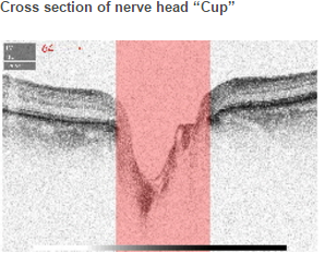
On presentation both eyes do have large cups. Full OCT scans were done of the back of the eye.
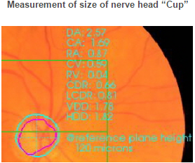
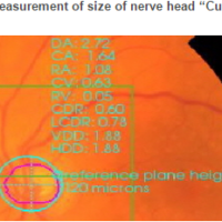
The Optic nerve does have some cupping and a notch can be seen when the software identifies the edge of the disc. Nerve thickness is shown to be less in the inferior temporal side of the Right nerve head and this is confirmed using the NEW macula central layer scans.
This lady was returned to the hospital for review with copies of all these scans and she was diagnosed with GLAUCOMA and is now being treated.
Without our GOLD scans we would have easily assumed that there was no change to her situation and as she had been cleared by the hospital eye service would have not been referred back for treatment.
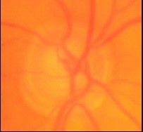
Right Eye
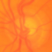
Left Eye
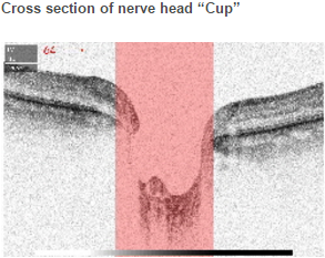
It can be seen that the “Cup” is quite large in both eyes.
There is a notch in the lower rim in the right eye at 7 o’clock which without the edges identified by the software is impossible to see. See blank pictures magnified below.
Both cups are large at 0.8 Disc Diameter being taken up by the cup horizontally.